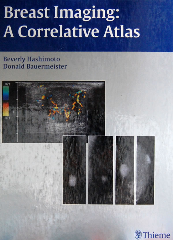
乳腺影像图谱Breast Imaging: A Correlative Atlas
 点此进入淘宝搜索页搜索
分類: 图书,进口原版书,医学 Medicine ,
作者: Beverly Hashimoto, Donald Bauermeister 著
出 版 社: Oversea Publishing House
出版时间: 2002-2-1字数:版次: 1页数: 536印刷时间:开本: 16开印次: 1纸张:I S B N : 9781588901095包装: 精装编辑推荐
作者简介:Beverly E. Flashimoto, M.D., F.A.C.R., is Section Head, Department of Ultrasound at Virginia Mason Medical Center and Clinical Assistant Professor of Radiology at the University of Washington, Seattle, Washington. Donald E. Bauermeister, M.D., is Chief of Pathology at Virginia Mason Medical Center and is Clinical Professor of Pathology at the University of Washington, Seattle, Washington.
内容简介
The book is targeted for breast imagers who wish to improve their interpretative skills by learning a pattern approach method to analyze and integrate mammographic and sonographic findings. One goal of the book is to emphasize the multidisciplinary nature of breast imaging utilizing sonography, magnetic resonance and various nuclear medicine techniques. Another goal is to demonstrate the importance of high-resolution sonography. The final objective of this book is to provide an atlas of a wide variety of pathological entities within the breast.
It includes coverage of normal anatomy and variants, technology and instrumentation, histology for selected cases, and management.
Two table of contents allow readers to access suspected diagnosis by either mmaography or ultrasound findings. The first enables readers to identify the histologic entities that create specific mammographic findings; the second aloows readers to study the lesions that produce sonographic abnormalities.
目录
Foreword
Preface
Acknowledgments
Dedication
List of Contributors
I Approach to Mammographic Analysis
Chapter 1 Approach to Mammographic Analysis
II Ultrasound Technique and Cross-Correlation with Mammography
Chapter2 Ultrasound Technique and Cross-Correlation with Mammography
III Circumscribed Masses
A Fat Containing Masses
B Medium or High Density Masses
IV Irregular Masses
A Benign Masses
B Malignant
V Calcifications
A Large linear
B Milk of Calcium
C Large Round
D Round Lucent Center
E Eggshell/Rim
F Coarse Popcorn-Fibroadenoma
G Dystrophic
H Small Round/Punctate
I Amorphous/Indistinct Microcalcifications
J Heterogeneous/Pleomorphic Microcalcifications
K Fine Linear/Branching Microcalcifications
VI Increased Density
A Generalized Increased Density
B Large Area Increased Density
C Focal Asymmetric Density
VII Architectural Distortion
A Peripheral Distortion
B Central Distortion
VIII Male Breast
A Benign
B Malignant
IX Postsurgical Findings
A Augmentation Mammoplasty Findings
B Reduction Mammoplasty
C Findings After Diagnostic or Therapeutic Procedures for Neoplasm
X Masses Poorly Identified Mammographically
A Patient Unable to Tolerate Mammogram
B Palpable Masses
C Mammogram Underestimates Tumor Size
D Mass in Unusual Locations
E Ductal Abnormalities
Index